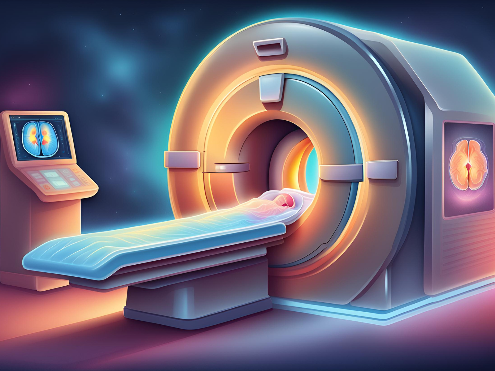
Magnetic Resonance Imaging (MRI) is a cornerstone in medical diagnostics, offering a non-invasive, powerful way to peer inside the human body in vivid detail. Unlike X-rays or CT scans, which use ionizing radiation, MRI employs magnetic fields and radio waves to create detailed images of organs and tissues. This post delves into how MRI works, its applications, and the advantages it brings to modern medicine.
How MRI Works?
At its core, MRI is based on the principles of nuclear magnetic resonance, a physical phenomenon in which nuclei in a magnetic field absorb and re-emit electromagnetic radiation. In the context of MRI, the body is placed inside a strong magnetic field, causing protons, particularly those in water molecules, to align with the field. When a radiofrequency current is then pulsed through the body, these aligned protons are perturbed from their position. As they relax back to their original alignment, they emit radio waves, which are detected and converted into images by the scanner.
The level of detail in these images is largely dependent on the magnetic field strength, measured in Tesla units. Clinical MRIs typically operate at 1.5 to 3.0 Tesla, but higher-end models used for research can go beyond 7 Tesla, offering even more precision and clarity.
Applications of MRI
MRI’s ability to differentiate between soft tissues makes it invaluable across various medical fields. Here are some key applications:
- Neurology: MRI is unrivaled in brain imaging, aiding in the diagnosis of conditions like strokes, tumors, and degenerative diseases such as Alzheimer’s.
- Orthopedics: It’s crucial for assessing bone, joint, and soft tissue conditions, including torn ligaments and cartilage.
- Cardiology: Cardiac MRI provides a detailed view of the heart and major vessels, assisting in the evaluation of heart structure and function.
- Oncology: MRI helps in detecting and staging various cancers, including those of the breast and prostate.
Advantages of MRI
- Safety: MRI does not expose patients to ionizing radiation, making it safer for repeated use, particularly in vulnerable populations such as pregnant women and children.
- Detail: It provides unparalleled detail of soft tissues, enhancing diagnostic accuracy.
- Versatility: Techniques like functional MRI (fMRI) can assess brain activity by measuring changes in blood flow, opening new avenues in neurological research.
Considerations and Limitations
Despite its advantages, MRI is not without limitations. The cost of MRI is significantly higher compared to other imaging techniques, which can limit accessibility. Patients with implanted medical devices, such as pacemakers, often cannot undergo MRI due to the strong magnetic field. Furthermore, the procedure can be time-consuming and requires the patient to remain still for extended periods, which can be challenging for some.
What is MRI Sequence?
An MRI sequence is a series of specific settings and timings of radiofrequency pulses and gradients that manipulate the behavior of hydrogen protons in the body’s tissues to create diverse contrast in the resulting images. Each sequence can emphasize certain tissue characteristics over others, making MRI incredibly versatile for diagnosing a wide range of conditions.
Key MRI Sequences and Their Applications
T1-Weighted (T1W) Sequences
- Overview: These sequences use a shorter repetition time (TR) and echo time (TE), producing images where fat appears bright and water appears dark.
- Clinical Use: T1W is essential for evaluating the brain’s anatomy, assessing musculoskeletal disorders, and post-contrast imaging to detect tumors and inflammation.
T2-Weighted (T2W) Sequences
- Overview: With longer TR and TE, these images make fluids appear bright, providing excellent contrast between different types of soft tissues.
- Clinical Use: T2W sequences are invaluable in diagnosing brain disorders, spinal pathologies, and joint abnormalities.
Fluid-Attenuated Inversion Recovery (FLAIR)
- Overview: FLAIR is a variation of T2W imaging that nullifies the signal from fluids like cerebrospinal fluid to better highlight lesions adjacent to fluid-filled areas.
- Clinical Use: It is predominantly used in brain imaging for detecting multiple sclerosis, strokes, and other cerebral abnormalities.
Diffusion-Weighted Imaging (DWI)
- Overview: DWI measures the diffusion of water molecules within tissue, producing images that can indicate cellular density and the integrity of cell membranes.
- Clinical Use: DWI is crucial for the early detection of strokes, characterizing tumors, and identifying abscesses.
Perfusion-Weighted Imaging (PWI)
- Overview: PWI assesses the flow of blood through tissues, often used to measure and visualize the amount of blood passing through various parts of the brain.
- Clinical Use: This sequence helps diagnose stroke, determine tumor malignancy levels, and assess brain tumors’ response to treatments.
Magnetic Resonance Angiography (MRA)
- Overview: MRA focuses on blood vessels, using MRI technology to detect vascular diseases without invasive catheterization.
- Clinical Use: It is used for evaluating aneurysms, vascular malformations, and stenoses.
Why MRI Sequences Are Crucial?
The flexibility in MRI sequences allows clinicians to tailor the scan according to the specific diagnostic needs, enhancing the ability to distinguish between normal and pathological tissues. This adaptability ensures that MRI is not only a tool for imaging but also a method for early diagnosis and monitoring of treatment response, particularly in complex cases like cancer or neurological disorders.
Conclusion
MRI remains a vital tool in medical diagnostics, blending advanced technology with patient safety to provide critical insights into the human body. As technology progresses, MRI’s capabilities will expand, continuing to revolutionize how doctors diagnose and treat disease, ensuring better patient outcomes.
The future of MRI holds promising advancements. With the development of stronger magnets, faster imaging techniques, and more sophisticated software that can extract even more detailed information from MRI data, the potential for even earlier disease detection and personalized medicine is on the horizon.